Brainbow
Never before has a brain been so beautiful. Jeff Lichtman and Joshua Sanes, researchers at the Harvard Brain Center, have created transgenic mice with fluorescent multicolored neurons. The photographs of the mouse brains that appear in the November 1, 2007 issue of Nature could be housed in the Museum of Modern Art or could be used to decorate Joseph’s technicolored dream coat. But it is not their colorful splendor that makes these genetically modified mice so amazing. It is their potential to revolutionize neurobiology that excites scientists like myself and has our neurons firing away, creating oodles of endorphins.
The mice created by a genetic strategy termed “brainbow” will have a similar effect on neuroscience as Google Earth had on cartography. Using a brainbow of colors, researchers will now be able to map the neural circuits of the brain. The individually colored neurons will help define the complex tangle of neurons that comprise the brain and nervous system. By creating a wiring diagram of the brain, researchers hope to help identify the defective wiring found in neurodegenerative diseases such as Altzheimer’s and Parkinson’s disease.
In the Brainbow mice, the Harvard researchers have introduced genetic machinery that randomly mixes green, cyan and yellow fluorescent proteins in individual neurons thereby creating a palette of ninety distinctive hues and colors. “The technique drives the cell to switch on fluorescent protein genes in neurons more or less at random,” says Jean Livet, the postdoctoral researcher responsible for most of the laboratory work that resulted in the Brainbow mice. “You can think of Brainbow almost like a slot machine in its generation of random outcomes.”
I can’t wait for a three dimensional multicolored atlas of the brain or a Google Brain site that is as beautiful as it is useful.
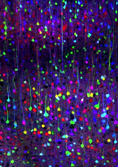
This is a photograph of the cerebral cortex. It’s hard to believe, but in non-living preserved brains, the outerlayers of this portion of the brain are gray, which is why the brain is sometimes called “gray matter”. (Confocal image by Tamily Weissman. Mouse by Jean Livet and Ryan Draft.)
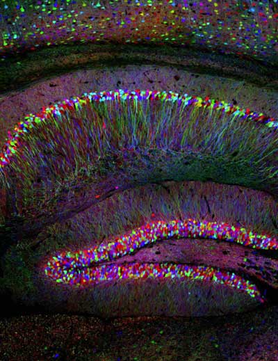
Another image of the cerebral cortex, which plays an important role in memory, perceptual awareness, thought and language. (Confocal image by Tamily Weissman. Mouse by Jean Livet and Ryan Draft.)
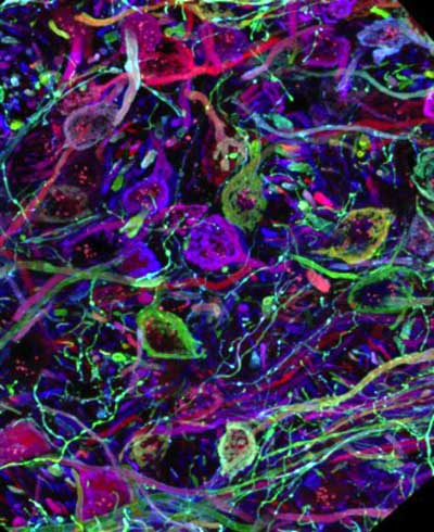
On this confocal microscopy image from the brain stem, tube-like structures are axons of the auditory pathway, which are forming hand-shaped synapses on other neurons (here unlabeled). (Confocal image by Jean Livet)