GRAB - Electron Tomography
Electron micrographs are colorless and so it is rather unexpected to find GFP being used in electron microscopy. But as this site shows uses for GFP can be found everywhere. GRAB (GFP Recognition After Bleaching) is a technique that uses the fact GFP generates oxygen radicals upon bleaching (i.e. the photochemical destruction of the chromophore) to locate GFP in electron microscopy. Markus Grabenbauer, who is now at the Max Planck Institute in Dortmund, came up with the idea to use the oxygen radicals that are generated during the GFP bleaching to photoxidize 3,3′-diaminobenzidine into an electron-dense solid that can be visualized by electron microscopy and electron tomography. The technique was first described in Nature Methods in November 2005. To show the utility of their method they fused a glycosylating enzyme to enhanced GFP and recorded electron tomograms of a Golgi stack containing GFP tagged enzyme, see the computer generated and color coded 3D model extracted from the GRAB tomogram.

A 3D model extracted from the tomogram of a Golgi stack containing the GFP tagged glycosylating enzyme (GalNAc-T2-GFP). The GalNAc-T2-GFP containing cisternae are colored green, vesicles containing GalNAc-T2-GFP are red, other vesicles are white and the endoplasmic reticulum is purple. A vitual section of the tomogram, showing the DAB solid precipitate, is set as the background image. (Photo courtesy of Markus Grabenbauer and Nature Methods)
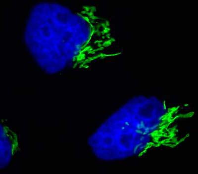
This is a fluorescence light microscopy image showing the GFP tagged GalNAC-T2 in the Golgi. It shows both microscopy techniques can be directly correlated: one for recording protein dynamics in vivo, and the other one to get higher resolution. (Photo courtesy of Markus Grabenbauer and Nature Methods)